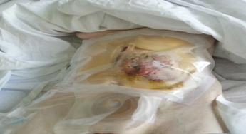
Management of Pseudomyxoma Peritonei

Management of Pseudomyxoma Peritonei

This case study looks at the management of Pseudomyxoma Peritonei using eakin Wound Pouches

By Deb Day, Stomal Therapy CNC Central Coast Local Health District, Australia

Patient background

A 47-year-old lady who had been diagnosed with Pseudomyxoma Peritionei 13 years earlier. She had multiple surgeries and a left-sided, high output ileostomy. The lady was very independent and lived at home on total parenteral nutrition (TPN) and intravenous fluids (IVF) via a Hickman’s Line.

Diagnosed with Pseudomyxoma Peritionei 13 years earlier.
Left-sided, high output ileostomy.


The lady presented to STN clinic in July 2014 and was referred back to her surgeon who diagnosed disease recurrence of Pseudomyxoma Peritonei. It was later decided that the lady would not have any further surgery.

Care Management Plan

The lady was managed under Palliative Care and continued to receive TPN, IVF and blood transfusions alongside the use of a syringe driver for pain relief.

The lady was managed under Palliative Care
Pouching the Pseudomyxoma Peritonei with eakin Wound Pouches™


Pouching the Pseudomyxoma Peritonei with eakin Wound Pouches was an appropriate choice as they provided comfort, odour control and containment of fluid from the wound. They also reduced the workload for the nurses providing care.

Outcome

Provides containment of fluid.

Provides comfort and odour control for the patient.

Reduced nurse workload.

No additional componentry or reliance on resources such as electricity.

Provided comfort, odour control and containment of fluid from the wound

One eakin Maxi Wound PouchTM solves the Problem

One Eakin Maxi Wound Pouch™ solves the Problem

This case study examines the role of one single Wound Pouch with Release Adhesive Remover in significantly improving patient comfort

Submitted by: Nicole Wienhold, RN, Abenraa Sygehus, Denmark.

Patient History

2010- 61 year old woman had a hysterectomy performed but surgery was complicated by a perforation of the sigmoid colon and a stoma was created.

Due to obesity the abdominal scar ruptured and was treated with VAC therapy.

2011- a parastomal hernia developed.

The wound was well managed by homecare nurse team.

The patient was worried about undergoing surgery to close the stoma and the wound. However, after careful consideration and evaluation, the decision was made to proceed with some plastic surgical assistance.

7 days post-operative, the wound dehisced. The lady developed severe sepsis and was admitted to the Intensive Care Unit.

She developed an enterocutaneous fistula.

Further surgery was carried out to close the stoma and the wound, but the fistula was not touched.

The lady has short bowel syndrome and has a central vein catheter to receive parenteral nutrition. She is NIDDM (Non-Insulin-Dependent Diabetes Mellitus) with unstable blood sugar.

Current Problems

Fig. 1 Coloplast pouches in place

The two Coloplast Fistula Pouches were found to be very rigid (Fig. 1), which meant when the patient moved, they leaked. Mobility is severely restricted due to frequent leakages.

Prior to removing the two Fistula Pouches, pain relief had to be administered to the patient. Morphine was given intravenously in advance of the wound dressing, as the pain was unbearable, even when using Welland Adhesive Remover Spray.


Care Management Plan

Fig. 3 Tracing the wound.
Fig. 2 Wound cleaned and uncovered.

Eakin Release Adhesive Remover Spray was recommended and even before the medication could take effect, the pouches were removed from her skin without any pain experienced. Fig. 2 shows the wound uncovered.

The patient’s large wound had deep cavities to the left and right. To avoid the accumulation of output (>2,000ml/24 hours), a Foley catheter was inserted into the fistula and the tube was allowed to drain out of the bung in the Wound Pouch into a drainage bag, which remained attached at all times.

Tracing guide used to draw the shape of the wound (Fig. 3).

Wound Pouch prepared.

One Eakin Maxi Wound Pouch was a perfect fit for this big wound.

The wound was picture framed (Fig. 4) with Eakin Cohesive® Skin Barriers cut in two long strips with small slits in one side to make it flexible around the wound.

Eakin Maxi Wound Pouch applied to the wound (Fig. 5).

"Fig.

"Fig.